Treatment of cerebral infarction

Treatment for cerebral infarction

KANAZAWA drip infusion treatment flow (Total of 5 outpatient treatments)

Inspection item


1)Blood test
2)electrocardiogram test
3)X-ray
4)ABI test
5)Platelet aggregation test
6)MRA, MRI

Additional information
・The number of drip infusion treatments is 5 times per course.
・Patients will undergo a blood test before the first drip infusion treatment, so please refrain from drinking anything other than water or non-caffeinated tea for 6 hours before starting treatment.
・A treatment plan will be determined during the first consultation.
・Patients consult with Dr. Kanazawa and decide whether to receive IV treatment for 5 consecutive days or once a week for a total of 5 times.
・A blood test will be conducted before the end of the fourth drip infusion treatment, so please do not drink anything other than water or non-caffeinated tea for 6 hours before the start of treatment.
・After the fifth drip infusion treatment, the doctor will conduct a detailed examination to confirm the effectiveness of the treatment and decide on future treatment plans.
・Inspections will be performed before the first drip infusion treatment and after the final (fifth) drip infusion treatment.
・Test items may change depending on the individual’s physical condition and symptoms.
・ On the day of your blood test, please do not drink anything other than water and decaffeinated tea for 6 hours before starting treatment.
・ For safety reasons, patients will not be able to receive Kanazawa drip infusion treatment if they do not undergo a preliminary examination.

Testing for MIBYO treatment

Tests for MIBYO treatment include the following

[1] medical examination
[2] EKG, chest radiograph, Holter EKG, echocardiogram
[3] Blood and biochemical tests, urine analysis, urine occult blood, urine ketones
[4] blood gas analysis
[5] Brain CT, brain MBRI, brain MRA
[6] Manifest artery MRA, cephalic artery echocardiography
[7] lumbar puncture
[8] brain waves
[9] cerebral angiography
[10] Cerebral artery lesions by 3D-CTA
[11] Measurement of cerebral circulation
  SPECT (Single Photon Emission Computed Tomography)
  PET (Positron Emission Tomography)
  Measurement of oxygen consumption with C15O2-15O2
  Zenon CT [12] Platelet aggregation capacity, blood coagulation capacity
[13] autonomic function
[14] thermography

*We do not always perform all tests. We perform a variety of tests using the most advanced technology of modern medicine according to the symptoms and conditions.

Vascular activation therapy has been established as a method to prevent cerebral infarction in its undiseased stage over a long period of half a century.
This method rejuvenates blood vessels and widens narrowed vessels.
It also improves blood flow. As a result, cerebral infarction can be prevented.

In the Kinsen infusion therapy, the right drug is used for each patient according to the medical examination. The IV therapy and lifestyle guidance will improve 6 symptoms.

Treatment of narrowing of arterial vessels: ・・・・・・・・

Widen narrowed arteries to improve blood flow

Reduce platelet pseudoaggregation・・・・・・・・

Blood vessels with atherosclerosis are easily damaged and platelets
platelets gather there and form blood clots.

LDL cholesterol, LDH (lactate dehydrogenase), 
Control of triglycerides, HbA1C, and Ua (uric acid). ・・・・・・・・

Factors that predispose to stroke can be improved through lifestyle changes and drugs.

Correction of neutrophilia, one of the white blood cells (reduce inflammation)・・・・・・・・

Reduce inflammation of blood vessel walls, which is a cause of cerebral infarction.

Correction of low hemoglobin・・・・・・・・

People who have cerebral infarction tend to be anemic, and this should be corrected by lifestyle and drugs.

Elimination of reactive oxygen species. ・・・・・・・・

People who have cerebral infarction tend to be anemic, and this is improved by lifestyle and drugs.

Correction of low hemoglobin・・・・・・・・

Removal of reactive oxygen species・・・・・・・・

The key is to improve these six symptoms.

From the past treatment, even the treatment to recover the pathology of the aftereffects of cerebral infarction and to prevent recurrence of the stroke has
improvement effect.

MRI and MRA are used to examine the brain itself and MRA to check the blood vessels.
The MRI and MRA are used to determine what diseases are present in the brain, which parts of the brain are damaged, and which blood vessels are affected.
Here are some of the main MRA images. The MRA images can be used as a reference to determine which parts of the cerebral blood vessels are clogged or stenosed, and to make changes in the brain that may result from treatment.
First of all, the MRA image shows what the arteries in the brain look like in the picture on the P-wing.

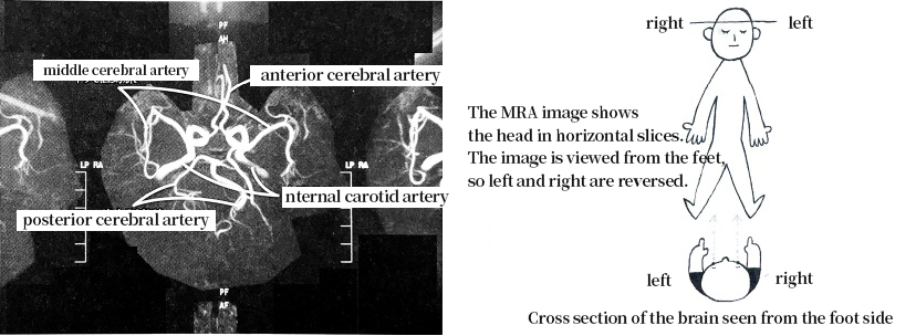
Normal human cerebral blood vessels

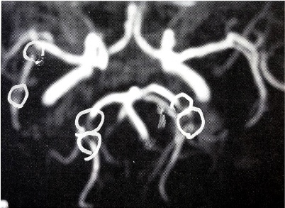
Cerebral vessels with stenosis

Don’t overlook the signs of cerebral infarction!

The shining white lines are blood vessels through which blood flows. If the white lines are connected as shown in the picture, the blood vessels are normal, but where stenosis has occurred, the areas that were white have become lighter or black. If a cerebral infarction occurs, there is no blood flow beyond that point, so the blood vessels do not show up.
By not overlooking these signs of stroke and starting treatment before the disease occurs, the risk can be avoided.

Example of treatment 69 years old male, Mr. A

He has lower extremity pain while walking and sometimes falls over due to dizziness.
He has started to feel a slight tremor in his hands.

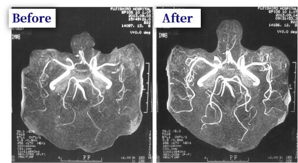
MRA showed bilateral middle cerebral artery stenosis and ischemic foci in the posterior cerebral artery. The patient was treated with intravenous infusion. After 10 days of intravenous infusion, the stenosis of the arterial vessels improved markedly and the patient's complaints almost disappeared.

Example of treatment 70 years old male, Mr. B

Example of improvement after stroke. Patient with right hemiparesis, dizziness and gait disturbance. MRA of the brain showed
The left middle cerebral artery is occluded, the left middle cerebral artery is stenotic, and the right posterior aorta is ischemic.

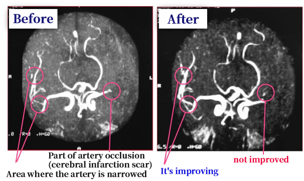
An example of a partially occluded area of an artery after stroke. After the same intravenous treatment as above. The occlusion of the left middle cerebral artery did not improve, but the stenosis of the other arteries improved markedly, and the patient's physical condition clearly improved.

The dead brain cells beyond the stroke site are not changed,
However, the narrowing of the area outside of the occlusion site is improved, and the symptoms of the brain beyond the occlusion site are restored.

As you know, blood vessels run throughout the body, and blood circulates to the ends of our fingertips.
The effects of unaffected therapy for cerebral infarction are, in fact, not limited to its effect on the brain.

Other Examples of Treatment

Example of treatment 70 years old male, Mr. C

Suspected of having latent pulmonary thrombotic symptoms, he was admitted to another hospital as pneumonia. After 2 months of treatment,
After 2 months of treatment, CT scan of the left side of the chest showed that there was still a shadow in the posterior and right middle lung lobe, and that she had difficulty breathing even after walking about 80 meters.
He had difficulty breathing after walking about 80 meters and had severe dynamic symptoms.

After treatment with intravenous infusion, most of the shadows disappeared as shown in the center of the picture, and in the right picture, most of the shadows disappeared,
The symptoms of dyspnea also improved. She no longer had difficulty breathing even after walking two kilometers to and from the hospital, and she no longer had severe dynamic symptoms.

Example of treatment 63 years old male, Mr. D

Mr. C has been smoking for 35 years. A case of obvious pulmonary fibrosis in the chest area.
He was admitted to the hospital as pneumonia without fever. She was treated with antibiotics but did not recover.

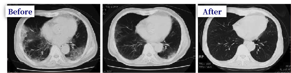
After treatment with intravenous gold therapy, the heterotopia almost disappeared and the symptoms disappeared.

Example of treatment 74 years old male, Mr. E

The patient came to the hospital complaining of walking pain, and as shown in the MRA, there was obvious vascular stenosis.

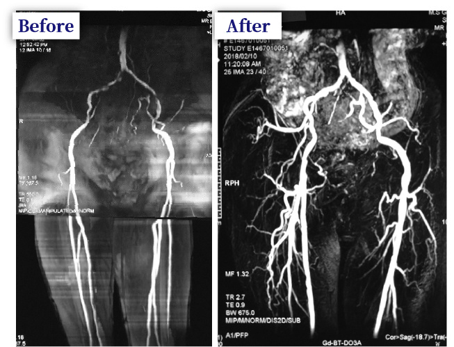
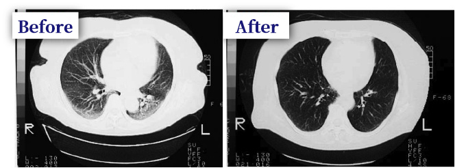
After the gold infusion therapy was administered, blood flow was clearly improved. He also said that most of his pain symptoms had been removed.